


 Katalogs


Case Provided by Prof Darko Božić, Zagreb, Croatia
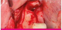
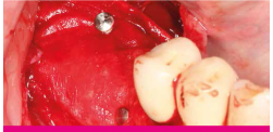
1. Patient with a distal mandibular edentulous ridge requiring implant placement:
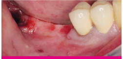
2. Flap elevation revealed significant loss of ridge height and width:
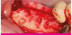
3. Edentulous ridge with significant loss of height and width:
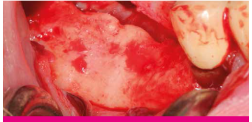
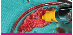
4. A small amount of autogenous bone was harvested leaving small cortical perforations:
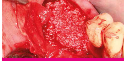
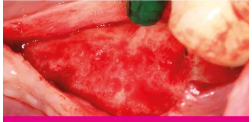
5. The autogenous bone was mixed with xenograft material saturated with xHyA:
6. Placement and adaptation of the graft mixture onto the recipient site:
7. The graft mixture was covered with a resorbable collagen membrane (SMARTBRANE) and fixed with pins.
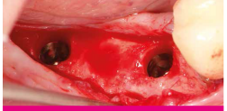
8. After 6 months. Significant gain of bone width with almost no residual graft particles visible
9. Implants of 4mm width were placed in the correct prosthetic positions:
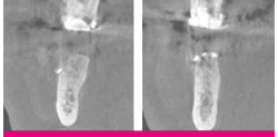
10. After 6 months. Cone beam computed tomography (CBCT) showing a significant amount of newly formed bone:

 Подписаться
 Купить в 1 клик
 Сравнение
 В избранное
 Недоступно

2020-2022 © SIA "Paradent - Baltic". Solutions for bone and tissue regeneration Republikas laukums 3 – 317, Riga, LV-1010, Latvia View on map |